A novel approach to studying the progression of tuberculosis (TB) from infection to disease has identified and treated people at increased risk of developing the disease that current methods of testing would not.
A collaboration between researchers from the University of Nottingham’s School of Biosciences and the National Institute for Health and Care Research (NIHR) Leicester Biomedical Research Centre (BRC) has resulted in the development of a novel method that could help with global efforts to reduce the spread of tuberculosis (TB).
Current test methods cannot identify those people exposed to TB that have the highest risk of developing active TB. The novel approach, published in the Lancet Microbe on 17 January, can specifically identify those patients, leading to faster treatment and better disease control.
This has been an exciting collaboration, and by bringing together different scientific disciplines we have gained new insights into the disease and this has allowed us to develop a new method to monitor how TB infections are progressing in individual patients.
Dr Pranabashis Haldar, Clinical Senior Lecturer in Respiratory Medicine at the University of Leicester and a Principal Investigator at the NIHR Leicester BRC, where the research was carried out, said: “Tuberculosis rates in the UK and around the world are not declining despite global efforts.
“TB is a bacterial disease that causes significant lung damage and can, without treatment, be fatal. It is spread in aerosol by inhaling droplets containing the bacteria. Most people that become infected live with the infection and remain well; however in a small proportion, the infection is not controlled and can progress to cause disease.
“Current tests of TB infection use either a skin test or a blood test, called an interferon gamma release assay (IGRA) to detect an immune response to the infection. However, these tests cannot distinguish between those that are at high or low risk of developing TB.
“An important research goal is to develop better tests that can identify the high risk group, so that we can provide more targeted treatment to prevent TB developing.”
In this study researchers used PET-CT, a highly sensitive form of imaging, as a novel way of looking at how the infection progresses, and to identify people at greater risk of developing the disease.
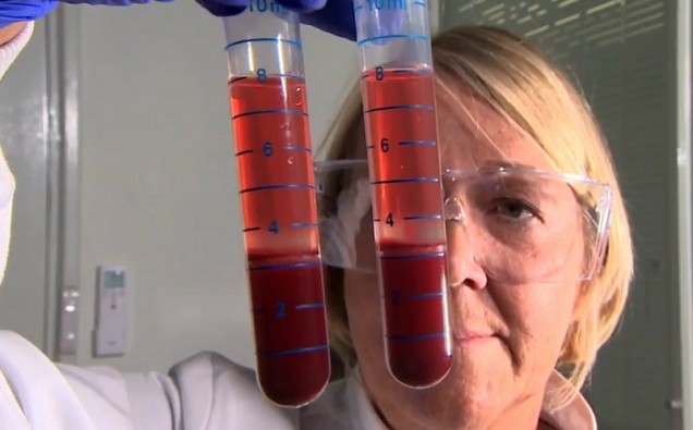
Additionally the team used a novel bacteriophage-based assay called Actiphage. This approach was developed by Professor Cath Rees from the University of Nottingham and PBD Biotech Ltd, a spin out company from the University of Nottingham.
Bacteriophages are viruses that infect bacterial cells and they are highly specific; with each phage preying on a single type of bacteria. The Actiphage assay uses a bacteriophage that attacks live TB bacteria; releasing the bacterial DNA which can then be detected by PCR. Using this approach, it is possible to detect very low levels of the bacterial DNA that cannot otherwise be detected using existing clinical tools.
This approach allowed the team to undertake a study evaluating a potential new blood test for identifying those at higher risk, without needing to recruit a large cohort, which can be challenging and very expensive.
Twenty adults traced back to households of people being treated for tuberculosis disease at University Hospitals of Leicester NHS Trust took part.
Participants underwent chest radiography and an IGRA to screen for TB infection. The research team then used two new methods of monitoring the progression of the disease over the following year: PET-CT imaging tools and a new blood test.
Dr Jee Whang Kim, a Clinical Research Fellow from the University of Leicester, who conducted the study said: “In PET-CT scans, patients are given fluorodeoxyglucose (FDG), a radiotracer which is similar to naturally occurring glucose (a type of sugar) that the body uses it in a similar way.
“By analysing the areas where the radiotracer is taken up, it’s possible to identify areas in the body where something might be going on.
“In this case, we were looking for evidence of metabolic activity associated with infection by the TB bacteria that cannot be seen using a chest x-ray or deduced by the blood tests used in routine clinical practice.
“In keeping with what is understood about the natural history of this infection, we found that the radiotracer activity tended to be taken up around the lungs, or in lymph nodes around the lungs. We then went on to perform a second PET-CT scan after 3 months to find out whether the infection was progressing or not. Where possible, we also took samples from the active sites to test for presence of the TB bacteria.”
The second novel aspect of the study was to use the Actiphage test to directly detect low levels of TB bacteria in the blood of patients with the infection.
“There is evidence of bacterial escape from where the primary infection occurs (the lungs) during progressive infection, and that escape might occur into the bloodstream,” added Dr Jee Whang Kim.
“Until now, studies have been limited by challenges of detecting low bacterial numbers.”
“We wanted to see whether this approach could identify metabolically active and replicating M tuberculosis in the blood of individuals who were otherwise completely well,” added Dr Jee Whang Kim.
We are just harnessing the natural ability of bacteriophage to find and break open the TB bacteria, and then the DNA that is released can be detected using conventional PCR tests. As well as its sensitivity, the other advantage of this method is that the PCR is only detecting DNA from live (viable) cells – if a dead TB cell is present, the bacteriophage won’t infect it so we know that a PCR detection event means that live cells are present in the blood sample.
In this study 20 TB contacts who were all asymptomatic with normal chest X-rays underwent a PET-CT baseline scan and, if it was positive and showed metabolic activity that could be sampled, they went on to have a bronchoscopy and sampling. If the baseline PET-CT scan did not show anything that could be sampled or if the sampling was negative for TB, they were monitored with a second PET-CT after three to four months.
Dr Haldar said: “Of the 20 contacts recruited to the study, one had a subtly abnormal chest radiograph that was picked up retrospectively. Using existing clinical tools and criteria, we can conclude that only this person may have been identified at routine contact screening to have—or be at higher risk of developing—tuberculosis.
“But using PET-CT we identified four people in whom the TB bacteria could be isolated from either the lung airway or PET-positive lymph nodes and two further people that had progressive changes after the second PET-CT scan. All six individuals were given TB treatment and in all of them, a further PET-CT scan 3 months after completing treatment showed resolving or completely resolved changes, further supporting our view that the PET-CT changes were caused by metabolically active tuberculosis infection.”
“We were also encouraged by the result of the Actiphage test,” added Dr Haldar
“We found a statistically significant association between a positive baseline Actiphage test and later being given treatment for high risk features of TB infection. Overall, Actiphage results were positive in 12 (60%) participants at baseline and positive in all six of the treated PET-CT- positive participants.
“Our results are exciting for two reasons. Firstly, they show that PET-CT could be an effective tool for identifying people with higher risk TB infection. This can help us to perform studies to develop new tests and evaluate new treatments, including vaccines more efficiently and at lower cost.
“Secondly, our findings suggest that TB bacteria are found in blood more often than has previously been thought and importantly, the presence of the bacteria in blood may be an indicator of uncontrolled or progressive TB infection.
“Based on our findings we propose that blood biomarkers aimed at detecting the bacteria, can complement existing biomarkers of the host immune response to enable better stratification of TB risk in those that have TB infection.”