Using a simple urine test alongside routine imaging for patients with adrenal masses could speed up adrenal cancer diagnosis, improving patient’s prognosis and reducing the need for invasive diagnostic procedures, a new multi-centre study published in The Lancet Diabetes & Endocrinology has found.
Imaging procedures, such as CT and MRI scans, are used in clinical practice with increasing frequency and often lead incidentally to the discovery of a nodule in the adrenal glands, detected on average in 5% of scans. These so-called adrenal incidentalomas are in the majority harmless, but once an adrenal mass has been discovered it is important to exclude adrenal cancer as well as adrenal hormone excess.
Prognosis for patients discovered to have an adrenal cortical carcinoma (ACC) – a cancerous adrenal mass – is poor, and a cure is only achievable through early detection and surgery. The incidental discovery of an adrenal mass often triggers additional scans to determine whether the mass is cancerous. However, recent studies have suggested that imaging tests have limited ability in establishing whether a mass is cancerous or benign.
Therefore, carrying out additional scans to characterise an adrenal mass increases costs, radiation exposure and anxiety in the patient, but mostly does not provide any more valuable information that could inform clinical management.
Led by experts from the University of Birmingham, a new multi-centre study, which is the first and the largest of its kind, has suggested that the addition of urine steroid metabolomics (USM) in the form of a simple urine test to detect the presence of excess adrenal steroid hormones – a key indicator of adrenal tumours – could speed up diagnosis and treatment for patients found to have an ACC and help eliminate the need for unnecessary surgery for patients with a harmless adrenal mass.
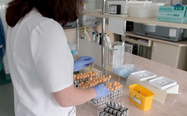
Over a six-year period, researchers studied more than 2000 patients with newly diagnosed adrenal tumours from 14 centres of the European Network for the Study of Adrenal Tumours (ENSAT). Patients collected a urine sample after being diagnosed and the researchers then analysed the types and amounts of adrenal steroids in the urine, with results automatically analysed by a machine learning based computer algorithm. Results showed that the urine test made fewer mistakes than imaging tests, which more frequently wrongly diagnosed ACC in a harmless adrenal nodule.
Professor Wiebke Arlt, Director of the Institute of Metabolism and Systems Research at the University of Birmingham and senior author of the study said: “Introduction of this new testing approach into routine clinical practice will enable faster diagnosis for those with cancerous adrenal masses. We hope that the results of this study could lead to significant decreases in patient burden and a reduction in healthcare costs, by not only reducing the numbers of unnecessary surgeries for those with benign masses, but also limiting the number of imaging procedures that are required.”
Dr Alice Sitch and Professor Jon Deeks, University of Birmingham diagnostic test specialists involved in the study, explained: “The study showed that the highest accuracy was provided when combining tumour size and imaging characteristics with the urine test, in particular when applying the urine test to patients with larger adrenal masses and suspicious looking imaging results.
“Following the initial scan that leads to the discovery of the adrenal mass, this combined test strategy would only have required further imaging in 488 (24.2%) of the study’s 2017 participants, who actually underwent 2737 scans prior to diagnostic decision.”
Irina Bancos, joint first author and Associate Professor of Endocrinology at the Mayo Clinic, Rochester, USA, said: “The findings of this study will feed into the next International guidelines on the management of adrenal tumours, and the implementation of the new test will hopefully improve the overall outlook for patients diagnosed with adrenal tumours.”
Angela Taylor, Research Fellow at the University of Birmingham and joint first author, explains: “This study shows the power of high-throughput steroid profiling by mass spectrometry, which we used to analyse the more than 2000 urine samples here in our Steroid Metabolome Analysis Core at the University of Birmingham.”
Michael Biehl, Professor of Computer Science at the University of Groningen, The Netherlands, said: “It is highly rewarding to see our transparent and interpretable algorithm validated in this large prospective study, which constitutes a superb example of truly interdisciplinary and international collaboration. This study paves the way to one of the first implementations of machine learning based classifiers in clinical practice.”


